Introduction
Acute alcohol intoxication is a common toxicological emergency, and severe poisoning may cause profound neurological impairment, respiratory depression, aspiration, coma, and death (1–3). Airway assessment is particularly challenging because reduced consciousness does not invariably correspond to ventilatory failure; decisions should integrate Glasgow Coma Scale (GCS), airway reflexes, respiratory effort, arterial blood gas findings, and the anticipated clinical course (3,4,8). Co-ingestion of benzodiazepines or opioids may intensify central nervous system depression. Benzodiazepines potentiate GABA-mediated sedation, whereas opioids directly suppress brainstem respiratory drive and reduce responsiveness to hypercapnia; combined exposure with alcohol may therefore increase the risk of hypoventilation, carbon dioxide retention, respiratory acidosis, and airway intervention (5–7). Although acute alcohol intoxication has been widely studied, few reports have simultaneously evaluated neurological status, arterial blood gas abnormalities, toxicological co-exposure, and airway management according to the class of co-ingested depressant within a single cohort of patients with alcohol-related coma. The present study was designed to address this gap. The aim of this study was to evaluate respiratory depression among patients presenting with alcohol-related coma and to investigate the impact of concomitant opioid or benzodiazepine exposure on respiratory compromise and clinical outcomes.
Co-intoxication with alcohol and opioids or benzodiazepines increases the risk of respiratory depression, hypercapnia, respiratory acidosis, and the need for endotracheal intubation.
The primary objective was to assess the association between mixed intoxication and the need for endotracheal intubation. Secondary objectives included the evaluation of arterial blood gas abnormalities, particularly hypercapnia and respiratory acidosis, as well as in-hospital mortality by type of co-ingested central nervous system depressant.
Materials and methods
Study Design and Setting
This retrospective observational cohort study was conducted at the Clinical Toxicology Service of the University Hospital Center “Mother Teresa” (QSUT), Tirana, Albania. Adult patients admitted with alcohol-related coma between January 2023 and December 2025 were retrospectively identified through hospital medical records. The study was designed to evaluate the impact of concomitant opioid or benzodiazepine exposure on respiratory depression, airway intervention, and short-term clinical outcomes in patients with severe acute alcohol intoxication.
Study Population
Medical records of consecutive adult patients (≥18 years) admitted during the study period were reviewed. Eligible patients had toxicologically confirmed acute alcohol intoxication associated with clinically significant impairment of consciousness requiring hospital admission and intensive monitoring.Alcohol-related coma was operationally defined as acute severe depression of consciousness predominantly attributable to ethanol after exclusion of alternative causes, including traumatic brain injury, stroke, hypoglycemia, sepsis, and primary neurological disease, on the basis of clinical evaluation and routine emergency investigations. A fixed GCS threshold was not required for inclusion because airway risk in poisoning depends on respiratory status, airway reflexes, aspiration risk, co-ingestion, and clinical trajectory in addition to GCS. Patients with incomplete medical records, unavailable arterial blood gas analysis, or missing toxicological data were excluded.
A total of 48 patients fulfilled the eligibility criteria and were included in the final analysis.
Exposure Classification
Patients were classified into three predefined exposure groups according to toxicological findings and clinical documentation: isolated alcohol intoxication, alcohol with concomitant benzodiazepine exposure, and alcohol with concomitant opioid exposure. Blood alcohol concentration (BAC) was measured on admission using standard hospital laboratory procedures. Identification of opioids and benzodiazepines was based on routine toxicological screening performed during emergency evaluation, together with the corresponding clinical findings.
Toxicological screening was performed on urine samples collected at hospital admission using qualitative immunoassay-based drugs-of-abuse assays on the Abbott ARCHITECT c8000 clinical chemistry platform. The screening panel included benzodiazepines and opiates. The manufacturer-defined qualitative cut-offs used by the laboratory were 300 ng/mL for opiates, expressed as morphine equivalents, and 300 ng/ mL for benzodiazepines, expressed as oxazepam equivalents. Confirmatory gas chromatography–mass spectrometry or liquid chromatography–tandem mass spectrometry was not routinely available. Results were therefore interpreted as evidence of exposure rather than quantitative drug concentration. Potential cross-reactivity, false-positive or false-negative findings, and limited detection of some synthetic opioids were recognized limitations of the screening methodology.
Clinical Assessment and Data Collection
Demographic characteristics, toxicological findings, clinical variables, respiratory parameters, and in-hospital outcomes were extracted from electronic medical records.
Demographic variables included age and sex. Neurological status at presentation was evaluated using the Glasgow Coma Scale (GCS), a validated instrument for assessing the level of consciousness in critically ill patients. Neurological assessment was performed immediately upon admission, before therapeutic interventions, whenever feasible.
Available records regarding pre-hospital or emergency department naloxone administration were also reviewed. Naloxone administration was documented in eight patients with opioid co-ingestion. The timing, dose, and immediate clinical response were not consistently recorded; naloxone was therefore summarized descriptively and was not included in inferential analyses.
Respiratory function was assessed by arterial blood gas analysis obtained at admission prior to definitive airway intervention whenever clinically possible. Arterial pH and partial pressure of arterial carbon dioxide (PaCO₂) were selected as objective markers of respiratory depression and ventilatory impairment because hypercapnia and respiratory acidosis represent the principal physiological consequences of central respiratory suppression during severe alcohol intoxication and mixed central nervous system depressant exposure.
Study Outcomes
The primary outcome of the study was the requirement for endotracheal intubation during hospitalization. Decisions regarding airway management were made by the treating emergency and intensive care physicians according to routine institutional practice, taking into consideration neurological status, airway protective reflexes, respiratory effort, arterial blood gas abnormalities, and the anticipated risk of respiratory deterioration rather than relying solely on the Glasgow Coma Scale, consistent with current evidence on airway management in intoxicated patients. Secondary outcomes included arterial blood gas abnormalities, particularly hypercapnia and respiratory acidosis, as well as in-hospital mortality. No predefined study protocol mandated intubation; consequently, this physician-dependent outcome may also reflect local practice variation and decision bias.
Statistical Analysis
Statistical analyses were performed using R statistical software (R Foundation for Statistical Computing, Vienna, Austria). Continuous variables were summarized as mean ± standard deviation. Categorical variables were summarized as frequencies and percentages. Distributional assumptions were assessed by graphical inspection and descriptive summary statistics. The distributions of admission GCS, PaCO₂, and arterial pH were additionally presented using boxplots, in which the horizontal line represents the median and the box represents the interquartile range.
Comparisons among the three intoxication groups were performed using one-way analysis of variance (ANOVA) for continuous variables and the chi-square test or Fisher’s exact test for categorical variables, as appropriate. Comparisons between intubated and non-intubated patients were performed using Student’s t-test for continuous variables and the chi-square or Fisher’s exact test for categorical variables.
To further investigate factors associated with endotracheal intubation, sequential Firth-penalized logistic regression models were constructed. Firth penalization was selected because of the relatively small sample size, sparse subgroup structure, and the possibility of quasi-complete separation. Initial models evaluated the association between intoxication group and airway intervention, whereas subsequent models incorporated blood alcohol concentration and neurological status (GCS). Covariates were restricted to a small number of clinically selected variables. Model discrimination was assessed using receiver operating characteristic (ROC) analysis and the apparent area under the ROC curve (AUC). Given the limited sample size and absence of internal or external validation, all regression and AUC analyses were considered exploratory and hypothesis-generating rather than confirmatory measures of predictive performance.
All statistical tests were two-sided, and a p-value <0.05 was considered statistically significant.
Results
Baseline Characteristics
A total of 48 patients with toxicologically confirmed alcohol-related coma were included in the study. Of these, 25 patients (52.1%) had isolated alcohol intoxication, 13 (27.1%) had concomitant benzodiazepine exposure, and 10 (20.8%) had concomitant opioid exposure.
Baseline demographic characteristics were comparable across the three groups. No statistically significant differences were observed in age, sex distribution, or blood alcohol concentration, indicating similar levels of alcohol exposure at presentation (Table 1).
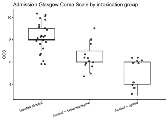
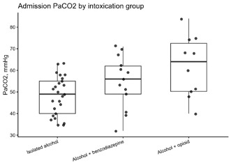
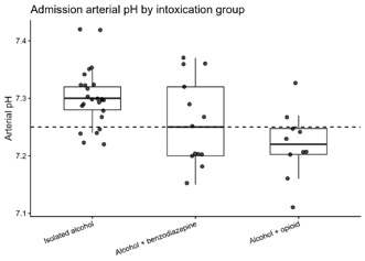
In contrast, significant differences were identified in neurological and respiratory parameters. Admission Glasgow Coma Scale (GCS) scores progressively decreased from patients with isolated alcohol intoxication to those with benzodiazepine and opioid co-ingestion (p<0.001). Similarly, arterial pH declined while PaCO₂ increased across exposure groups, demonstrating progressively more severe respiratory compromise in patients with mixed intoxication. Patients with alcohol–opioid co-ingestion exhibited the lowest GCS scores, the highest PaCO₂ values, and the lowest arterial pH values, indicating the greatest degree of respiratory depression at presentation (Figures 2–4).
Distribution of admission Glasgow Coma Scale (GCS) scores in patients with isolated alcohol intoxication, alcohol–benzodiazepine co-ingestion, and alcohol–opioid co-ingestion. Boxes represent the interquartile range (IQR), the horizontal line indicates the median, whiskers extend to 1.5 × IQR, and individual points represent individual patients.
Distribution of admission arterial PaCO₂ values among the three intoxication groups. Boxes represent the interquartile range (IQR), the horizontal line indicates the median, whiskers extend to 1.5 × IQR, and individual points represent individual patients.
Distribution of admission arterial pH values among the three intoxication groups. Boxes represent the interquartile range (IQR), the horizontal line indicates the median, whiskers extend to 1.5 × IQR, and individual points represent individual patients. The dashed horizontal line indicates the predefined threshold for severe respiratory acidosis (pH 7.25).
Clinical Outcomes
Clinical outcomes according to intoxication group are summarized in Table 2.
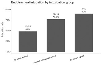
Overall, 31 of 48 patients (64.6%) required endotracheal intubation during hospitalization. The frequency of airway intervention increased progressively according to the pattern of intoxication, ranging from isolated alcohol intoxication to alcohol–benzodiazepine and alcohol–opioid co-ingestion (Figure 1). Patients with opioid co-ingestion demonstrated the highest intubation rate, consistent with the greater severity of neurological impairment and ventilatory dysfunction observed at admission.
Percentage of patients requiring endotracheal intubation in the three intoxication groups: isolated alcohol intoxication (12/25, 48.0%), alcohol–benzodiazepine co-ingestion (10/13, 76.9%), and alcohol–opioid co-ingestion (9/10, 90.0%).
Overall, in-hospital mortality was low (2/48, 4.2%). Both deaths occurred in patients with mixed intoxication, whereas no deaths were recorded among patients with isolated alcohol intoxication. Although this difference did not reach statistical significance, the observed pattern suggests greater clinical severity in patients exposed to additional central nervous system depressants.
Naloxone administration was documented in eight patients, all in the alcohol–opioid co-ingestion group. Because the timing, dosage, and immediate clinical response were inconsistently documented, no inferential analysis of naloxone response was performed.
Respiratory Depression According to Intoxication Pattern
Objective markers of respiratory function demonstrated a clear gradient of increasing physiological impairment across the three exposure groups.
Admission GCS decreased progressively in parallel with worsening arterial blood gas abnormalities. Patients with concomitant opioid exposure presented with the greatest degree of hypercapnia and respiratory acidosis, whereas patients with isolated alcohol intoxication showed the least severe physiological abnormalities. Patients with alcohol–benzodiazepine co-ingestion demonstrated intermediate findings between these two groups.
The distribution of individual GCS, PaCO₂, and arterial pH values is illustrated in Figures 2–4. These data demonstrate a consistent association between mixed intoxication, impaired consciousness, progressive carbon dioxide retention, and worsening respiratory acidosis.
Factors Associated with Endotracheal Intubation
To further characterize factors associated with airway intervention, patients were stratified according to intubation status (Table 3).
Compared with non-intubated patients, those requiring endotracheal intubation presented with significantly lower admission GCS scores, lower arterial pH values, and substantially higher PaCO₂ concentrations. In contrast, age, sex distribution, and blood alcohol concentration did not differ significantly between the two groups.
These findings indicate that objective measures of neurological depression and ventilatory impairment were more closely associated with the need for endotracheal intubation than demographic characteristics or blood alcohol concentration alone.
Multivariable Analysis of Endotracheal Intubation
Exploratory Firth-penalized logistic regression was performed to examine factors associated with endotracheal intubation.
Sequential adjustment demonstrated that neurological impairment and respiratory dysfunction provided incremental prognostic information beyond intoxication pattern and blood alcohol concentration (Table 4).
After incorporating the Glasgow Coma Scale into the model, the apparent AUC was 0.947 (95% CI 0.891–1.000). This estimate represents unvalidated within-cohort discrimination and may be inflated by overfitting because of the limited sample size.
These exploratory findings require confirmation in larger prospective cohorts with internal and external validation.
Discussion
This retrospective study suggests that concomitant central nervous system depressants, particularly opioids, identify patients with more severe neurological and respiratory compromise despite comparable blood alcohol concentrations. The principal clinical interpretation is not that intoxication pattern alone determines the need for intubation, but that suspected co-exposure should prompt integrated assessment of consciousness, ventilatory status, arterial blood gas abnormalities, airway reflexes, and the anticipated clinical course. Respiratory depression remains one of the most clinically important complications of severe acute alcohol intoxication and a major determinant of morbidity in patients presenting with impaired consciousness. Ethanol depresses central nervous system activity through multiple neurochemical mechanisms, thereby impairing airway-protective reflexes, reducing arousal, and attenuating ventilatory responsiveness (2). As intoxication progresses, these effects may lead to hypoventilation, carbon dioxide retention, and respiratory acidosis. In the present study, mixed intoxication was associated with significantly greater respiratory compromise than isolated alcohol intoxication, as reflected by higher PaCO₂ levels and lower arterial pH values. These observations support the concept that respiratory dysfunction is a principal mechanism linking mixed intoxication to adverse clinical outcomes. Conzelmann et al. demonstrated that reduced consciousness and increasing clinical severity were closely associated with aspiration risk in patients with severe acute alcohol intoxication, emphasizing the importance of comprehensive airway assessment (4).
Airway management should be guided by the overall clinical picture, including consciousness, ventilatory status, airway reflexes, and arterial blood gas abnormalities, rather than neurological status or blood alcohol concentration alone.
Similarly, Sauter et al. reported that decisions regarding endotracheal intubation should not rely solely on neurological status but should incorporate the overall clinical presentation (3). Our findings reinforce this multidimensional approach, showing that objective indicators of ventilatory impairment closely paralleled increasing clinical severity across the exposure groups. Together, these observations suggest that arterial blood gas analysis provides clinically relevant information beyond neurological assessment alone and may facilitate early recognition of patients at increased risk of respiratory deterioration and the need for advanced airway management. This integrated approach is consistent with contemporary recommendations for the evaluation of poisoned patients, in which neurological examination, respiratory physiology, and toxicological assessment are considered complementary rather than isolated determinants of airway management decisions (12,16).
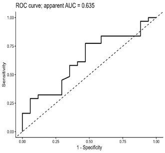
Figure 5. Receiver operating characteristic (ROC) curve of the unadjusted logistic regression model (Model A) evaluating the association between opioid co-ingestion and endotracheal intubation. The apparent area under the curve (AUC) was 0.635, indicating modest discriminative ability prior to adjustment for clinical severity.
One of the principal findings of the present study was the substantially greater respiratory compromise observed among patients with concomitant opioid exposure. Compared with both isolated alcohol intoxication and alcohol–benzodiazepine co-ingestion, patients in the opioid group exhibited the highest PaCO₂ values, the lowest arterial pH levels, and the greatest frequency of endotracheal intubation. Although the retrospective nature of the study precludes causal inference, these findings are biologically plausible and consistent with the established mechanisms of opioid-induced respiratory depression. Activation of μ-opioid receptors within brainstem respiratory centers suppresses respiratory drive, decreases ventilatory responsiveness to hypercapnia, and promotes progressive carbon dioxide retention (6).
When combined with alcohol, which independently depresses central nervous system function, these pharmacological effects may become synergistic, resulting in more profound impairment of consciousness and respiratory function than either agent alone (5). Our observations are consistent with previous reports demonstrating increased morbidity and mortality associated with concurrent exposure to opioids, alcohol, and other sedative agents (5,7). Tori et al. reported that alcohol and benzodiazepines were frequently involved in opioid-related overdose deaths, highlighting the clinical relevance of mixed intoxication in routine practice (7). Although mortality was uncommon in our cohort, all in-hospital deaths occurred in patients with mixed intoxication, supporting the concept that concomitant exposure to additional central nervous system depressants is associated with greater clinical severity. Moreover, the progressive decline in GCS, accompanied by increasing hypercapnia and worsening respiratory acidosis observed in the opioid group, suggests a continuum of physiological deterioration preceding the need for advanced airway intervention. These findings emphasize the importance of early recognition of opioid co-ingestion, close respiratory monitoring, and timely arterial blood gas assessment in patients presenting with alcohol-related coma, as these objective physiological markers may facilitate early identification of individuals at increased risk of respiratory decompensation and endotracheal intubation. Although the respiratory effects of benzodiazepines are generally less pronounced than those associated with opioids, their concomitant use with alcohol remains clinically important. Benzodiazepines potentiate γ-aminobutyric acid (GABA)-mediated inhibitory neurotransmission, the same neurochemical pathway through which ethanol exerts many of its sedative effects (14,15).
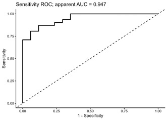
Figure 6. Receiver operating characteristic (ROC) curve of the severity-adjusted logistic regression model.
As a result, concurrent exposure may produce enhanced central nervous system depression, greater impairment of consciousness, and reduced protective airway reflexes compared with alcohol intoxication alone. This pharmacodynamic interaction represents a well-recognized mechanism underlying the increased clinical severity frequently observed in mixed alcohol-benzodiazepine intoxication. In the present cohort, patients with alcohol-benzodiazepine exposure demonstrated greater neurological and respiratory impairment than those presenting with isolated alcohol intoxication, although the magnitude of these abnormalities remained less pronounced than that observed among patients with opioid co-ingestion. This finding is consistent with current pharmacological understanding. While benzodiazepines may contribute substantially to sedation and impairment of airway protective mechanisms, they generally exert a less profound direct effect on ventilatory drive than opioids (5,15).
Consequently, respiratory compromise in alcohol-benzodiazepine intoxication is often mediated through a combination of reduced consciousness, impaired airway protection, and secondary hypoventilation rather than severe primary respiratory suppression. From a clinical perspective, these observations emphasize that alcohol-benzodiazepine intoxication should not be regarded as a benign condition. Even in the absence of profound hypercapnia, patients may remain vulnerable to aspiration, airway obstruction, and progressive deterioration of mental status. Previous studies have highlighted the risks associated with concurrent exposure to alcohol and benzodiazepines, particularly when additional sedative agents are present (5,7,12).
Our findings support the need for careful monitoring of this patient population and reinforce the importance of considering co-ingested substances when assessing the severity of alcohol-related coma. The management of the airway in patients presenting with alcohol-related coma remains one of the most challenging aspects of emergency and critical care practice. Although endotracheal intubation is frequently performed to protect the airway and ensure adequate ventilation, the optimal criteria for intervention remain controversial. Historically, a Glasgow Coma Scale score below 8 has often been regarded as an indication of the need for airway protection. However, accumulating evidence suggests that neurological assessment alone may not adequately identify patients at risk of respiratory deterioration (3,9,11).
Intubated patients had significantly lower arterial pH and higher PaCO2, highlighting the importance of objective ventilatory abnormalities in airway decision-making.
Our findings support a more comprehensive approach to airway evaluation. Patients who underwent endotracheal intubation had significantly lower GCS scores, lower arterial pH values, and substantially higher PaCO₂ levels than nonintubated patients. Importantly, these observations suggest that objective evidence of ventilatory impairment contributed to clinical decision-making beyond the degree of impaired consciousness alone. Similar conclusions have been reported by Sauter et al., who observed that intubation practices in acute alcohol intoxication were influenced by the overall clinical presentation rather than GCS alone (3). Because intubation was determined by treating physicians rather than by a study protocol, the observed associations may partly reflect local practice patterns and decision bias.
Likewise, Donald and colleagues identified several clinical factors beyond neurological status that were associated with the need for rapid sequence intubation in poisoned patients (9). Collectively, previous studies support the concept that airway management in acute poisoning should be guided by the overall clinical presentation rather than by a single neurological threshold (3,4,9,11,12). Conzelmann et al. demonstrated a close relationship between impaired consciousness and aspiration risk in severe alcohol intoxication (4), whereas Burket et al. emphasized the complexity of airway management decisions and the limitations of relying on a single clinical variable when determining the need for intubation (11). More recently, Freund et al. showed that carefully selected poisoned patients with impaired consciousness could be managed without immediate intubation under close clinical supervision, further supporting an individualized approach to airway management (12). Our findings are consistent with this evidence and suggest that airway management in alcohol-related coma should integrate neurological examination, respiratory assessment, and overall clinical status. In this context, arterial blood gas analysis appears to be a valuable adjunct, providing objective evidence of ventilatory failure and facilitating early identification of patients at increased risk of respiratory decompensation. Such a multidimensional assessment strategy is increasingly recommended in contemporary toxicology practice because no single clinical variable adequately captures the complexity of poisoning-related respiratory failure (12,16).
Early clinical assessment should integrate neurological status, respiratory physiology, and toxicological exposure to better identify patients at risk of clinical deterioration.
The multivariable analyses further reinforce the importance of objective clinical assessment. Adjustment for blood alcohol concentration did not materially alter the association between intoxication pattern and endotracheal intubation, indicating that ethanol concentration alone provides limited information regarding immediate clinical risk. In contrast, admission Glasgow Coma Scale score remained the strongest independent correlate of airway intervention, supporting the concept that clinical decision-making should be guided primarily by neurological and physiological status rather than blood alcohol concentration alone. These observations are consistent with previous reports demonstrating that the clinical manifestations of alcohol intoxication are determined not only by ethanol exposure but also by concomitant central nervous system depressants and individual physiological susceptibility. Accordingly, early assessment that integrates a neurological examination and arterial blood gas analysis may provide greater clinical value than reliance on ethanol concentration alone.
The apparent improvement in model discrimination after incorporating clinical severity variables should be interpreted with particular caution. With only 48 patients and small exposure subgroups, the AUC of 0.947 may substantially overestimate performance because of model overfitting. Formal internal validation was not feasible, and the reported discrimination therefore represents apparent rather than validated model performance. These analyses are exploratory and hypothesis-generating and require confirmation in larger prospective multicentre studies before clinical implementation.
Clinical Implications and Future Directions
The findings of the present study have several practical implications for clinicians managing patients with alcohol-related coma. First, they emphasize the importance of actively identifying concomitant opioid or benzodiazepine exposure during the initial assessment of patients presenting with impaired consciousness, as mixed intoxication was consistently associated with greater neurological and respiratory compromise. Second, the observed association between hypercapnia, respiratory acidosis, and endotracheal intubation supports the routine integration of respiratory assessment, including arterial blood gas analysis, into the evaluation of severely intoxicated patients. Finally, these findings reinforce the need for individualized airway management strategies that integrate neurological examination, respiratory physiology, and overall clinical status rather than relying exclusively on consciousness scores. Future prospective multicentre studies involving larger patient populations are warranted to externally validate these findings and further define the role of physiological markers such as PaCO₂ and arterial pH in predicting clinically relevant outcomes in alcohol-related coma.
Interpretation of Comparative Literature
As summarized in Table 5, the available evidence consistently supports the importance of respiratory dysfunction and individualized airway management in patients with severe alcohol intoxication and poisoning. The findings of the present study are broadly consistent with previous reports while extending current knowledge by demonstrating a clear gradient of neurological impairment, hypercapnia, respiratory acidosis, and endotracheal intubation across isolated alcohol intoxication, alcohol–benzodiazepine co-ingestion, and alcohol–opioid co-ingestion. By integrating toxicological exposure, neurological assessment, arterial blood gas analysis, and airway intervention within a single cohort, the present study provides additional evidence that objective respiratory and neurological parameters may offer greater clinical value than blood alcohol concentration alone when assessing patients with alcohol-related coma.
Limitations
This study has several limitations that should be considered when interpreting the findings. First, its retrospective single-center design introduces the possibility of selection and referral bias, as patients admitted to a tertiary intensive care unit may not be representative of the broader population with acute alcohol intoxication. Second, the relatively small sample size, particularly within the mixed-intoxication subgroups, limited statistical power and constrained the complexity of multivariable analyses despite the use of Firth-penalized logistic regression. Third, toxicological confirmation relied primarily on qualitative urine immunoassays, which may yield false-positive or false-negative results, are affected by cross-reactivity and sampling time, and may not detect some synthetic opioids. Fourth, endotracheal intubation was a physician-dependent outcome based on institutional practice; variability in clinical judgment and decision bias cannot be excluded. Fifth, several potentially relevant confounders, including chronic respiratory disease, chronic alcohol dependence, body mass index, aspiration events, and pre-hospital interventions, were not consistently available. The timing, dose, and immediate clinical response to naloxone were also incompletely documented. Finally, the prediction models were not internally or externally validated; the apparent AUC of 0.947 may be optimistic because of overfitting.Accordingly, the findings should be interpreted as hypothesis-generating and require external validation before routine clinical application.
Conclusions
In this retrospective cohort of patients with alcohol-related coma, concomitant opioid and benzodiazepine exposure was associated with greater neurological and respiratory impairment than isolated alcohol intoxication, with opioid co-ingestion showing the most severe physiological profile. Because of the small sample and exploratory analyses, these associations should not be interpreted as a validated prediction rule. The findings support early toxicological screening and prompt arterial blood gas analysis in patients presenting with alcohol-related coma to facilitate individualized airway management, while confirmation in larger prospective multicentre studies is required.
Conflict of Interest
The authors declare no conflicts of interest relevant to the content of this study.
Funding
This research received no external funding.
Ethics Approval
This retrospective observational study was conducted using anonymized clinical data obtained from the Clinical Toxicology Service of the University Hospital Center “Mother Teresa” (QSUT), Tirana, Albania. The study was performed in accordance with the ethical principles of the Declaration of Helsinki and was approved by the appropriate institutional ethics committee. Due to the retrospective nature of the study and the use of anonymized patient data, the requirement for informed consent was waived.
References
- World Health Organization. Global status report on alcohol and health and treatment of substance use disorders 2024. Geneva: World Health Organization; 2024.
- Mirijello A, Sestito L, Antonelli M, Gasbarrini A, Addolorato G. Identification and management of acute alcohol intoxication. Eur J Intern Med. 2023; 108:1-8. doi: 10.1016/j.ejim.2022.08.013.
- Sauter TC, Rönz K, Hirschi T, Lehmann B, Exadaktylos AK, Hautz WE. Intubation in acute alcohol intoxications at the emergency department. Scand J Trauma Resusc Emerg Med. 2020;28(1):11. doi:10.1186/s13049-020-0707-2.
- Conzelmann M, Hoidis A, Bruckner T, Popp E, Koschny R. Aspiration risk in relation to Glasgow Coma Scale score and clinical parameters in patients with severe acute alcohol intoxication: a single-centre retrospective study. BMJ Open. 2021;11(10). doi:10.1136/bmjopen-2021-053619.
- Gudin JA, Mogali S, Jones JD, Comer SD. Risks, management, and monitoring of combination opioid, benzodiazepine, and/or alcohol use. Postgrad Med. 2013;125(4):115-130. doi:10.3810/pgm.2013.07.2684.
- Algera MH, Kamp J, van der Schrier R, van Velzen M, Niesters M, Aarts L et al. Opioid-induced respiratory depression in humans: a review of pharmacokinetic-pharmacodynamic modelling of reversal. Br J Anaesth. 2019;122(6). doi: 10.1016/j.bja.2019.03.009.
- Tori ME, Larochelle MR, Naimi TS. Alcohol or benzodiazepine co-involvement with opioid overdose deaths in the United States, 1999-2017. JAMA Netw Open. 2020;3(4). doi:10.1001/jamanetworkopen.2020.2361.
- Brandenburg R, Brinkman S, de Keizer NF, Kesecioglu J, Meulenbelt J, de Lange DW. The need for ICU admission in intoxicated patients: a prediction model. Clin Toxicol (Phila). 2017;55(1):4-11. doi:10.1080/15563650.2016.1222616.
- Donald C, Duncan R, Thakore S. Predictors of the need for rapid sequence intubation in the poisoned patient with reduced Glasgow Coma Score. Emerg Med J. 2009;26(7):510-512. doi:10.1136/emj.2008.064998.
- Rönz K, Exadaktylos AK, Sauter TC. Predictors of high resource consumption in alcohol-intoxicated patients in the emergency department. Int J Environ Res Public Health. 2020;17(11):4122. doi:10.3390/ijerph17114122.
- Burket GA, Horowitz BZ, Hendrickson RG, Beauchamp GA. Endotracheal intubation in the pharmaceutical-poisoned patient: a narrative review of the literature. J Med Toxicol. 2021;17(1):61-69. doi:10.1007/s13181-020-00779-3.
- Freund Y,Viglino D, Cachanado M, Cassard C, Montassier E, Douay B, et al. Effect of noninvasive airway management of comatose patients with acute poisoning: a randomized clinical trial. JAMA. 2023;330(23):2267–74. doi:10.1001/jama.2023.24391.
- Strayer RJ, Friedman BW, Haroz R, Ketcham E, Klein L, LaPietra AM et al. Emergency department management of patients with alcohol intoxication, alcohol withdrawal, and alcohol use disorder: a white paper prepared for the American Academy of Emergency Medicine. J Emerg Med. 2023;64(4):517-540. doi:10.1016/j.jemermed.2023.01.010.
- Gaudreault P, Guay J, Thivierge RL, Verdy I. Benzodiazepine poisoning: clinical and pharmacological considerations and treatment. Drug Saf. 1991;6(4):247-265. doi:10.2165/00002018-199106040-00003.
- Tanaka E. Toxicological interactions between alcohol and benzodiazepines. J Toxicol Clin Toxicol. 2002;40(1):69-75. doi:10.1081/CLT-120002886.
- Azharuddin S, Ogbebor O, Shuster M, Smith B, Arshad H, Cheema T. Toxicological emergencies. Crit Care Nurs Q. 2023;46(1):82-99. doi:10.1097/CNQ.0000000000000439.