Introduction
Group A beta-hemolytic streptococcus (Streptococcus pyogenes) is a Gram-positive bacterium most commonly associated with upper respiratory tract infections, particularly pharyngitis, as well as skin and soft tissue infections.Although most infections are mild and self-limiting, invasive group A streptococcal (iGAS) infections remain an important cause of morbidity and mortality worldwide (1,2).
Clinical manifestations of invasive group A streptococcal infection include bacteremia, pneumonia, pleural infections, necrotizing fasciitis, streptococcal toxic shock syndrome (STSS), and multiple organ dysfunction syndrome (MODS) (2,3). Severe disease is associated with several virulence factors, including the M protein and streptococcal pyrogenic exotoxins, which act as superantigens and may trigger an exaggerated systemic inflammatory response, endothelial injury, coagulopathy, and multiple organ dysfunction (3). In recent years, an increase in the incidence of invasive group A streptococcal infections has been reported across several European countries and other developed regions, further emphasizing the importance of early recognition and timely treatment (4,5). Invasive infections may occur in individuals of all ages and with variable risk factors (3).
We report a case of rapidly progressive invasive Streptococcus pyogenes infection complicated by pleural empyema, pericardial effusion, sepsis, and multiple organ dysfunction syndrome in a previously healthy young adult.
Invasive Streptococcus pyogenes infection may rapidly progress from uncomplicated pharyngitis to sepsis with multiple organ dysfunction syndrome, even in previously healthy adults.
Case report
A previously healthy 28-year-old man with a history of penicillin allergy and active smoking presented to the Emergency Department with a four-day history of fever, sore throat, and vomiting. He had been treated with oral azithromycin in the outpatient setting for presumed streptococcal pharyngitis. One day before admission, he developed pleuritic pain in the left hemithorax radiating to the left shoulder.
On presentation, the patient was fully alert and afebrile (36.5 °C), but markedly tachypneic (respiratory rate 51 breaths/ min) and tachycardic (heart rate 130 beats/min), with a blood pressure of 135/75 mmHg and peripheral oxygen saturation of 96% on room air. Physical examination revealed enlarged tonsils without visible exudate. Pulmonary auscultation demonstrated markedly diminished breath sounds over the left lower lung field.
Laboratory investigations revealed a pronounced systemic inflammatory response characterized by markedly elevated inflammatory markers and lactate levels, thrombocytopenia, acute kidney injury, and coagulation abnormalities. During hospitalization, thrombocytopenia and leukocytosis transiently worsened before inflammatory markers, renal function, and liver enzyme levels gradually normalized. The temporal changes in the main laboratory parameters are presented in Table 1.
Radiological evaluation revealed a left-sided pleural effusion with compressive atelectasis of the adjacent lung parenchyma and findings consistent with pleural empyema, together with a small pericardial effusion without hemodynamic compromise. The chest radiograph obtained on admission is shown in Figure 1.
Blood cultures yielded Streptococcus pyogenes. According to the antimicrobial susceptibility testing, the isolate was susceptible to the empirically initiated antimicrobial therapy, which was therefore continued. Based on the clinical presentation and laboratory and imaging findings, the patient was admitted to the intensive care unit with a diagnosis of sepsis complicated by pleural empyema (pleural fluid was obtained for biochemical and microbiological analysis), pericarditis, acute kidney injury, and sepsis-induced coagulopathy. The NEWS2 score on admission was 5, indicating an intermediate risk of clinical deterioration. Empirical intravenous antimicrobial therapy with meropenem and clindamycin was initiated together with supportive treatment and low-molecular-weight heparin. Partial drainage of the pleural space was performed. Because of the markedly elevated D-dimer levels, pleuritic chest pain, and tachypnea, pulmonary embolism was initially considered in the differential diagnosis but was excluded by computed tomography pulmonary angiography. Deep vein thrombosis was excluded by duplex ultrasonography of the lower-extremity veins.
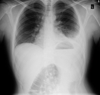
Figure 1. Chest radiograph on admission
During hospitalization, inflammatory markers gradually declined, renal function recovered, and the platelet count normalized. After 24 days of hospitalization, the patient was discharged home afebrile, without respiratory symptoms, and in good clinical condition. At discharge, elevated D-dimer levels persisted, consistent with the recent septic state.
Discussion
This case illustrates the rapid progression from acute pharyngitis to sepsis with multiple organ dysfunction in a previously healthy young adult. The patient fulfilled the Sepsis-3 criteria for sepsis, presenting with acute kidney injury and coagulation abnormalities. These findings represent organ dysfunction secondary to sepsis and reflect the well-established pathophysiological mechanisms of invasive group A streptococcal infection, including systemic inflammatory response and activation of the coagulation cascade (2,5).
A particularly noteworthy feature of this case was the simultaneous involvement of the pleura and pericardium. Pleural empyema is a rare but well-recognized complication of invasive group A streptococcal infection, whereas pericardial involvement is considerably less common and has mainly been described in isolated case reports (6,7). In our patient, imaging studies demonstrated pleural empyema, while echocardiography revealed a small circumferential pericardial effusion without hemodynamic significance.
Early recognition, prompt antimicrobial therapy, and timely source control are essential for favorable clinical outcomes.
Management of invasive group A streptococcal infection is based on early administration of appropriate antimicrobial therapy and prompt source control. In addition to beta-lactam antibiotics, clindamycin plays an important role because of its ability to inhibit streptococcal toxin production and is therefore recommended in severe invasive disease (2). In the present case, early recognition of sepsis, prompt initiation of antimicrobial therapy, and partial drainage of the pleural empyema resulted in complete clinical recovery.
Conclusion
This case demonstrates that invasive group A streptococcal infection may rapidly progress from apparently uncomplicated pharyngitis to sepsis with multiple organ dysfunction syndrome, even in previously healthy individuals. Early recognition of invasive disease, timely diagnostic evaluation, and prompt initiation of appropriate treatment are essential for achieving a favorable clinical outcome.
Reference
- Carapetis JR, Steer AC, Mulholland EK, Weber M. The global burden of group A streptococcal diseases. Lancet Infect Dis. 2005;5(11):685–94. doi:10.1016/S1473-3099(05)70267-X
- Walker MJ, Barnett TC, McArthur JD, Cole JN, Gillen CM, Henningham A et al. Disease Manifestations and Pathogenic Mechanisms of Group A Streptococcus. Clin Microbiol Rev. 2014;27(2):264–301. doi:10.1128/CMR.00101-13
- Mercadante S, Ficari A, Romani L, De Luca M, Tripiciano C, Chiurchiù S et al. The Thousand Faces of Invasive Group A Streptococcal Infections: Update on Epidemiology, Symptoms, and Therapy. Children. 2024;11(4):383. doi:10.3390/children11040383
- Johannesen TB, Munkstrup C, Edslev SM, Baig S, Nielsen S, Funk T et al. Increase in invasive group A streptococcal infections and emergence of novel, rapidly expanding sub-lineage of the virulent Streptococcus pyogenes M1 clone, Denmark, 2023. Eurosurveillance. 2023;28(26). doi:10.2807/1560-7917.ES.2023.28.26.2300291
- Brouwer S, Rivera-Hernandez T, Curren BF, Harbison-Price N, De Oliveira DMP, Jespersen MG et al. Pathogenesis, epidemiology and control of Group A Streptococcus infection. Nat Rev Microbiol. 2023;21(7):431–47. doi:10.1038/s41579-023-00865-7
- Leung CCD, Fong PY, Chan YH, Ho MY, Yeung YC. Two Cases of Group A Streptococcus-Induced Right Empyema: Rare Occurrences in Adult Medicine. Cureus. 2024;16(9):e68920. doi: 10.7759/cureus.68920
- Pemira SM, Tolan RW. Invasive Group A Streptococcus Infection Presenting as Purulent Pericarditis With Multiple Splenic Abscesses: Case Report and Literature Review. Clin Pediatr (Phila). 2012;51(5):436–41. doi:10.1177/0009922811430345