Introduction
Trismus is a clinical condition defined by a limited ability to open the mouth, arising from either functional disturbances or structural alterations within the masticatory system. While it is commonly regarded as a symptom rather than a distinct disease, it represents a clinically significant sign that may reflect underlying local or systemic disorders (1). This condition can substantially impair health-related quality of life (QoL), contributing to temporomandibular dysfunction (TMD), restricted food intake, muscle pain, and increased muscular tension (2).
Trismus is a multifactorial condition affecting mouth opening and function
In both dental and broader medical settings, identification of trismus is crucial, as it can compromise essential activities such as chewing, speaking, and maintaining oral hygiene. Furthermore, it presents a notable challenge in airway management, particularly during endotracheal intubation (3). In response to these risks, various professional organizations have established protocols and guidelines aimed at enhancing the safety and effectiveness of airway management (1–3).
Methodology
This manuscript is presented as a narrative review synthesizing both general information on trismus and evidence-based recommendations for managing the difficult airway. We conducted a comprehensive literature review using multiple databases, including PubMed, MEDLINE, and Google Scholar. Search terms included combinations of “difficult airway,”“ICU intubation,” and“trismus”. Boolean operators (AND, OR) were used to expand or narrow results based on thematic relevance. We included clinical practice guidelines (e.g., American Society of Anesthesiologists [ASA] 2022 (1) and Difficult Airway Society [DAS] 2025 (3)), randomized controlled trials, observational studies, meta-analyses, and expert consensus statements relevant to adult airway management in critical care and trismus.
Etiology
The etiology of trismus is diverse and multifactorial. Odontogenic infections are among the most frequent etiologies, especially those originating in the mandibular molar region, where inflammatory processes may spread to adjacent masticatory muscles. Trauma, including mandibular fractures or soft tissue injuries, may also result in restricted mouth opening due to pain, edema, or mechanical obstruction. Furthermore, TMD are a common contributing factor, with joint dysfunction or internal derangements playing a significant role in the development of trismus.
Complication of Infection
Dental infection must be suspected in patients with acute trismus, as severe dental infections involving the masticatory muscles often cause trismus (6). Such infections have the potential to spread into adjacent fascial spaces of the head and neck, occasionally leading to serious and life-threatening complications, including cervical cellulitis and mediastinitis (7). Among dental causes, pericoronitis is particularly commonly associated with the development of trismus (8). Examples of infections from non-odontogenic sources that may trigger trismus include tonsillitis, parotid abscess, tetanus, meningitis, and brain abscess (4).
Complication of Local Anesthesia
Trismus may occur as a complication following an inferior alveolar nerve block. During the administration of this block, the masticatory muscles, mainly the medial pterygoid muscle, can be accidentally penetrated, resulting in pain-related trismus. Stretching of the affected muscle provokes pain, triggering a reflex contraction that restricts mandibular movement (4).Additionally, the formation of an intramuscular hematoma may contribute to the development of trismus. Damage to the inferior alveolar vessels can result in bleeding within the pterygomandibular space, further limiting jaw mobility (4). When trismus develops within two to three days after the procedure, it is most commonly associated with infection along the needle pathway (5).
Odontogenic infections are a leading cause of acute trismus cases
Complication of Dental Extractions
Pain, limited mouth opening, and postoperative swelling are among the most frequently observed complications following the surgical removal of mandibular third molars, which influence the patient’s QoL in the postoperative period (6). These manifestations are primarily the result of the inflammatory response to surgical trauma and are short-term. The transient jaw stiffness usually peaks on the 2nd day and resolves by the end of the 1st week (7).
Sign of Temporomandibular Disorders (TMD)
Temporomandibular disorders (TMDs) encompass pain and dysfunction of the temporomandibular joint (TMJ) and the associated masticatory muscles. They are commonly present with facial and preauricular pain, joint sounds during function, and restricted mandibular movement. Consequently, patients presenting with reduced mouth opening often have an underlying TMD. Among the various subtypes, myofascial pain and disc displacement without reduction may contribute to the development of trismus (8).
Sign of Malignancy and/or complication of Head and Neck Cancer Treatment
Trismus is rarely the initial manifestation of malignancy; however, it is frequently observed in patients with head and neck cancer (9). Reported prevalence varies widely in the literature, ranging from 0% to 100%, depending on cancer location and extent (10). Further diagnostic evaluation, including radiographic imaging and specialist referral, should be considered when trismus is accompanied by the following clinical features (11):
- Maximal mouth opening less than 15 millimeters (mm)
- Absence of joint clicking history
- Pain of non-myofascial origin (e.g., neuralgia)
- Presence of lymphadenopathy
Trismus significantly complicates airway management in emergency care
Trismus is also a frequent complication of head and neck cancer and its treatment. Recent studies report an incidence of approximately 38% to 42% among affected patients (12,13). Radiotherapy may induce fibrosis of the temporomandibular joint or masticatory muscles, resulting in restricted mouth opening. The risk of trismus is particularly increased when the medial pterygoid muscle lies within the radiation field. In addition, surgical management of head and neck malignancies may lead to scarring of the masticatory musculature, further contributing to mandibular mobility (14).
In patients who develop trismus following oncological treatment, it is essential to distinguish whether it represents a treatment-related complication or an early sign of cancer recurrence (10).
Diagnosis, clinical presentation and management
Diagnosis of trismus is primarily established through clinical evaluation, including measurement of maximal mouth opening and assessment of accompanying symptoms.A comprehensive medical and dental history is essential for identifying the underlying etiology. In cases where structural abnormalities, trauma, or neoplastic conditions are suspected, additional imaging modalities such as computed tomography (CT) or magnetic resonance imaging (MRI) may be indicated (15). Clinically, trismus is characterized by a reduced interincisal distance, typically below 35 mm, whereas normal mouth opening ranges from 35 to 45 mm, with males generally exhibiting slightly greater values (approximately 40–60 mm, average 50 mm) (16).
Patients commonly report pain, stiffness, and difficulty performing everyday oral functions, including eating, speaking, and maintaining oral hygiene. In more advanced cases, nutritional intake and overall QoL may be significantly affected. The underlying cause largely determines management, with conservative treatment approaches preferred initially. These include physiotherapy with jaw-opening exercises, thermotherapy, and pharmacological interventions such as nonsteroidal anti-inflammatory drugs or muscle relaxants (9). In the presence of infection, appropriate antibiotic therapy and, when necessary, surgical drainage are required (7). Chronic or severe cases, particularly those associated with fibrosis, may necessitate more advanced treatments, including jaw mobilization devices or surgical intervention. If left untreated, trismus can result in multiple complications, such as impaired oral hygiene, increased risk of dental disease, speech difficulties, and nutritional deficiencies. Therefore, early diagnosis and appropriate management are essential to prevent long-term functional impairment (14).
Difficulties in Emergency and Clinical Care
Trismus is a well-recognized contributor to difficult airway management and represents a significant clinical challenge that requires a structured, well-prepared approach. Limitation of mouth opening directly interferes with standard airway techniques, complicating both ventilation and intubation. In severe cases, trismus can render conventional approaches, such as orotracheal intubation or the use of supraglottic airway devices, impossible. As a result, airway management in these patients remains one of the most demanding aspects of emergency and intensive care practice, where rapid decision-making and technical expertise are critical to preventing adverse outcomes.
Early diagnosis and imaging are key in identifying underlying etiology
Over the past decade, several professional associations have developed comprehensive guidelines to improve the safety and success of difficult airway management. The American Society of Anesthesiologists updated its difficult airway guidelines in 2022, emphasizing early recognition of airway difficulty, pre-procedural planning, and the integration of modern tools such as video laryngoscopy (1). Similarly, the Difficult Airway Society (DAS) published its 2025 guidelines (3), which provide a clear, stepwise algorithm for managing unanticipated difficult intubation in adults, progressing from initial intubation attempts to emergency front-of-neck access if required. In the intensive care setting, the Intensive Care Society, in collaboration with DAS, issued guidelines in 2018 that address the unique challenges of critically ill patients, particularly highlighting the importance of achieving first-pass success and minimizing hypoxia (17). Comparable recommendations have also been published by the French Society of Anesthesia and Intensive Care, as well as broader critical care organizations such as the American College of Chest Physicians and the American Thoracic Society, all reinforcing the need for preparedness and standardized approaches (18). Despite these advances, unanticipated difficult airways continue to occur and are associated with serious complications, including hypoxia, brain injury, and death. Trismus significantly contributes to this risk by impairing bag-mask ventilation and limiting access to the oropharynx. In situations where nasal intubation is contraindicated and mouth opening is severely restricted, alternative strategies such as submental intubation or awake tracheostomy may be required. However, these techniques are invasive and carry a higher risk of complications, including infection, bleeding, fistula formation, tracheal injury, voice changes, and cosmetic deformities, when compared with standard oral intubation. For this reason, emphasis is increasingly placed on early identification of potential airway difficulty, careful planning, and the use of less invasive techniques whenever feasible (19). Predicting difficult airway scenarios remains inherently challenging. Therefore, clinicians must maintain a high level of vigilance and be prepared for unexpected difficulties in all settings. When uncertainty exists, cautious direct laryngoscopy under topical anesthesia or minimal sedation may be performed to assess the feasibility of intubation.Awake tracheal intubation, in particular, has been shown to have a high success rate and favorable safety profile, yet it remains underutilized despite being strongly recommended in cases of anticipated difficult airway (2,20).
Multidisciplinary approach improves outcomes in trismus management
Among the various etiological factors, odontogenic infections are one of the most common causes of trismus. These infections can rapidly spread to adjacent fascial spaces, increasing the risk of airway compromise and systemic complications. Prompt and effective management is therefore essential. The cornerstone of treatment is incision and drainage of the abscess, which relieves pressure, reduces swelling, and eliminates the primary source of infection, often resulting in immediate clinical improvement.Adjunctive antibiotic therapy is indicated in cases of systemic involvement, such as fever, diffuse swelling, or immunocompromised status (21,22).
In selected clinical scenarios, alternative airway techniques may provide additional options. One such method is retromolar intubation (Figure 1), which utilizes the anatomical space located posterior to the last molar, bounded by the mandibular ramus and maxillary tuberosity. This space is present in all individuals and can be accessed by retracting the angle of the mouth. Although often described as a distinct technique, many reports refer to standard intubation followed by repositioning of the endotracheal tube into the retromolar space. This approach preserves dental occlusion and offers particular advantages in maxillofacial, orthognathic, oncologic, and trauma surgeries, where surgical access and occlusal alignment are critical (23,24).
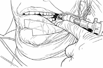
Figure 1. Retromolar intubation
Overall, the management of trismus-related difficult airway requires a multidisciplinary approach involving anesthesiologists, surgeons, and nursing staff. Continuous education, simulation-based training, and familiarity with evolving airway technologies are essential components of improving patient outcomes. Given the potentially life-threatening consequences of airway compromise, a proactive, well-coordinated strategy remains the cornerstone of safe and effective management.
Conclusion
Trismus is a multifactorial clinical condition with significant implications for both dental and medical practice. Its diverse etiology requires thorough clinical evaluation to identify the underlying cause and guide appropriate management. Beyond its impact on oral function and quality of life, trismus represents a critical concern in emergency and intensive care settings due to its association with difficult airway management. Despite advances in airway guidelines and techniques, it continues to pose a substantial challenge, particularly in unanticipated scenarios. Early recognition, accurate diagnosis, and timely intervention are essential to prevent complications and improve patient outcomes. A multidisciplinary approach, supported by adherence to evidence-based guidelines and ongoing clinician training, remains fundamental in ensuring safe and effective management of patients with trismus.
Acknowledgment
The authors would like to thank all individuals and institutions who contributed to this study.
References
- Apfelbaum JL, Hagberg CA, Connis RT, Abdelmalak BB, Agarkar M, Dutton RP et al. 2022 American Society of Anesthesiologists Practice Guidelines for Management of the Difficult Airway. Anesthesiology. 2022;136(1):31–81. doi:10.1097/ALN.0000000000004002.
- Martins MP, Ortenzi AV, Perin D, Quintas GCS, Malito ML, Carvalho VH. Recommendations from the Brazilian Society of Anesthesiology (SBA) for difficult airway management in adults. Braz J Anesthesiol. 2023;74(1):744477. doi:10.1016/j.bjane.2023.12.001.
- DAS guidelines for management of unanticipated difficult intubation in adults. Difficult Airway Society [Internet]. [cited 2026 Apr 10]. Available from: https://das.uk.com/guidelines/das_intubation_guidelines/
- Haas DA. Localized complications from local anesthesia. J Calif Dent Assoc. 1998;26(9):677–82.
- Ogle OE, Mahjoubi G. Local anesthesia: agents, techniques, and complications. Dent Clin North Am. 2012;56(1):133–48, ix. doi:10.1016/j.cden.2011.08.003.
- Bui CH, Seldin EB, Dodson TB. Types, frequencies, and risk factors for complications after third molar extraction. J Oral Maxillofac Surg Off J Am Assoc Oral Maxillofac Surg. 2003;61(12):1379–89. doi:10.1016/j.joms.2003.04.001.
- Balakrishnan G, Narendar R, Kavin T, Venkataraman S, Gokulanathan S. Incidence of Trismus in Transalveolar Extraction of Lower Third Molar. J Pharm Bioallied Sci. 2017;9(Suppl 1):S222–7. doi:10.4103/jpbs.JPBS_161_17.
- List T, Jensen RH. Temporomandibular disorders: Old ideas and new concepts. Cephalalgia Int J Headache. 2017;37(7):692–704. doi:10.1177/0333102416686302.
- Abboud WA, Hassin-Baer S, Alon EE, Gluck I, Dobriyan A, Amit U et al. Restricted Mouth Opening in Head and Neck Cancer: Etiology, Prevention, and Treatment. JCO Oncol Pract. 2020;16(10):643–53. doi:10.1200/OP.20.00266.
- Rapidis AD, Dijkstra PU, Roodenburg JLN, Rodrigo JP, Rinaldo A, Strojan P, et al. Trismus in patients with head and neck cancer: etiopathogenesis, diagnosis and management. Clin Otolaryngol Off J ENT-UK Off J Neth Soc Oto-Rhino-Laryngol Cervico-Facial Surg. 2015;40(6):516–26. doi:10.1111/coa.12488.
- Beddis HP, Davies SJ, Budenberg A, Horner K, Pemberton MN. Temporomandibular disorders, trismus and malignancy: development of a checklist to improve patient safety. Br Dent J. 2014;217(7):351–5. doi:10.1038/sj.bdj.2014.862.
- Shao CH, Chiang CC, Huang TW. Exercise therapy for cancer treatment-induced trismus in patients with head and neck cancer: A systematic review and meta-analysis of randomized controlled trials. Radiother Oncol J Eur Soc Ther Radiol Oncol. 2020;151:249–55. doi:10.1016/j.radonc.2020.08.024.
- Cohen EEW, LaMonte SJ, Erb NL, Beckman KL, Sadeghi N, Hutcheson KA, et al. American Cancer Society Head and Neck Cancer Survivorship Care Guideline. CA Cancer J Clin. 2016;66(3):203–39. doi:10.3322/caac.21343.
- Epstein JB, Thariat J, Bensadoun RJ, Barasch A, Murphy BA, Kolnick L et al. Oral complications of cancer and cancer therapy: from cancer treatment to survivorship. CA Cancer J Clin. 2012;62(6):400–22. doi:10.3322/caac.21157.
- Dhanrajani PJ, Jonaidel O. Trismus: aetiology, differential diagnosis and treatment. Dent Update. 2002;29(2):88–92, 94. doi:10.12968/denu.2002.29.2.88.
- Kerawala C, Newlands C. Oral and maxillofacial surgery. Oxford: Oxford University Press; 2010.
- Higgs A, McGrath BA, Goddard C, Rangasami J, Suntharalingam G, Gale R et al. Guidelines for the management of tracheal intubation in critically ill adults. Br J Anaesth. 2018;120(2):323–52. doi:10.1016/j.bja.2017.10.021.
- Mosier JM, Sakles JC, Law JA, Brown CA, Brindley PG. Tracheal Intubation in the Critically Ill. Where We Came from and Where We Should Go. Am J Respir Crit Care Med. 2020;201(7):775–88. doi:10.1164/rccm.201908-1636CI.
- Liaqat T, Amjad MA, Cherian SV. Difficult Airway Management in the Intensive Care Unit: A Narrative Review of Algorithms and Strategies. J Clin Med. 2025;14(14):4930. doi:10.3390/jcm14144930.
- Apfelbaum JL, Hagberg CA, Connis RT, Abdelmalak BB, Agarkar M, Dutton RP et al. 2022 American Society of Anesthesiologists Practice Guidelines for Management of the Difficult Airway. Anesthesiology. 2022 J;136(1):31–81. doi:10.1097/ALN.0000000000004002.
- Ryan P, McMahon G. Severe dental infections in the emergency department. Eur J Emerg Med Off J Eur Soc Emerg Med. 2012;19(4):208–13. doi:10.1097/MEJ.0b013e32834ddb68.
- Kinzer S, Pfeiffer J, Becker S, Ridder GJ. Severe deep neck space infections and mediastinitis of odontogenic origin: clinical relevance and implications for diagnosis and treatment. Acta Otolaryngol (Stockh). 2009;129(1):62–70. doi:10.1080/00016480802008181.
- Togioka BM, Pinkert V, Geltzeiler M, Chen EM. Retromolar intubation for severe trismus: a less invasive alternative to tracheostomy and submental intubation. BMJ Case Rep. 2021;14(1):e239817. doi:10.1136/bcr-2020-239817.
- Martinez-Lage JL, Eslava JM, Cebrecos AI, Marcos O. Retromolar intubation. J Oral Maxillofac Surg. 1998;56(3):302–5. doi:10.1016/S0278-2391(98)90103-3.